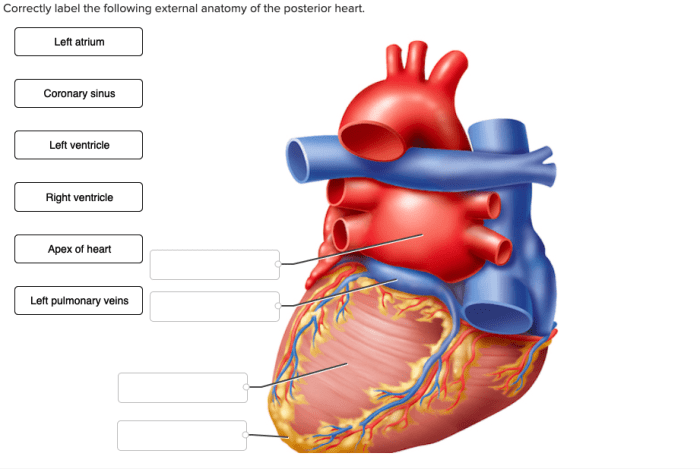
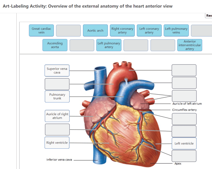
Correctly label the following external anatomy of the posterior heart – Correctly labeling the external anatomy of the posterior heart is crucial for understanding its structure and function. This guide provides a comprehensive overview of the posterior heart’s external anatomy, including its location, orientation, and major structures.
External Anatomy of the Posterior Heart
The posterior heart, located on the dorsal side of the heart, exhibits a distinct anatomy. Its orientation is such that the apex of the heart points towards the left, while the base is directed towards the right. The posterior surface of the heart is primarily composed of the left atrium, the right atrium, and the coronary sulcus.
The left atrium, positioned on the left side of the posterior heart, is separated from the right atrium by the posterior interatrial sulcus. The right atrium, located on the right side of the posterior heart, is adjacent to the left atrium and is separated from the left ventricle by the coronary sulcus.
Sulci and Grooves: Correctly Label The Following External Anatomy Of The Posterior Heart
The posterior heart is characterized by several sulci and grooves that serve important functional roles. The coronary sulcus, the most prominent groove, encircles the heart and separates the atria from the ventricles. Within the coronary sulcus lies the coronary sinus, which collects deoxygenated blood from the heart muscle.
The posterior interventricular sulcus, located between the left and right ventricles, extends from the coronary sulcus to the apex of the heart. It accommodates the posterior interventricular artery.
Coronary Vessels

The posterior heart is supplied by branches of the coronary arteries. The right coronary artery (RCA) gives rise to the posterior descending artery (PDA), which supplies the posterior interventricular sulcus and the posterior wall of the left ventricle. The left circumflex artery (LCX) contributes to the posterior interventricular sulcus and supplies the left atrium and the posterior wall of the right ventricle.
Atrioventricular Grooves

The atrioventricular grooves are located at the junction of the atria and ventricles. The right atrioventricular groove accommodates the right coronary artery, while the left atrioventricular groove contains the left coronary artery.
Within the atrioventricular grooves, the coronary sinus, a large venous channel, receives deoxygenated blood from the heart muscle via its tributaries, including the great cardiac vein, middle cardiac vein, and small cardiac vein.
Posterior Interatrial Sulcus

The posterior interatrial sulcus is a groove that separates the left and right atria on the posterior surface of the heart. It extends from the coronary sulcus to the apex of the heart and accommodates the interatrial branch of the left coronary artery.
The posterior interatrial sulcus plays a crucial role in the electrical conduction system of the heart. The atrioventricular node, located within the sulcus, receives electrical impulses from the sinoatrial node and transmits them to the bundle of His, initiating ventricular contraction.
FAQ Guide
What is the posterior heart?
The posterior heart refers to the back surface of the heart.
What are the major structures visible on the posterior surface of the heart?
The major structures visible on the posterior surface of the heart include the left atrium, right atrium, coronary sulcus, and posterior interventricular sulcus.
What is the significance of the sulci and grooves on the posterior heart?
The sulci and grooves on the posterior heart mark the boundaries between different heart chambers and facilitate the passage of blood vessels and nerves.